Researchers find new pathway to regulate immune response, control diseases


Researchers find new pathway to regulate immune response, control diseases

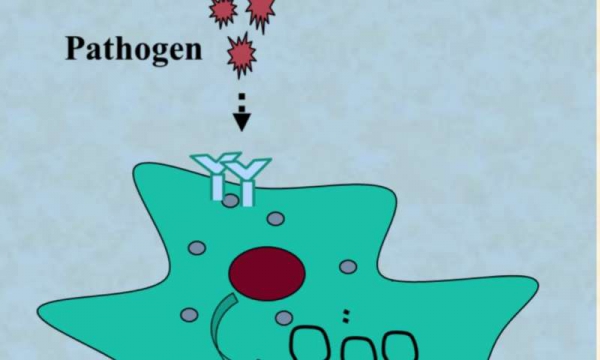
Non-coding RNA (ncRNA) regulates cytokines expression, immune response, and inflammation. Credit: University of Texas at Arlington

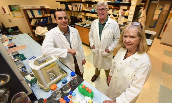
Researchers at The University of Texas at Arlington have found a potential new pathway to regulate immune response and potentially control inflammatory diseases of the central nervous system such as meningitis and sepsis.

“We need to know what turns on inflammatory response to bacterial infection to be able to modulate the process,” said Subhrangsu Mandal, the UTA associate professor of chemistry who led the research.

“If we can do so, we can control inflammatory diseases of the central nervous system that have been hard to treat up to now, such as sepsis and meningitis, as well as cancer and muscular dystrophy, which can also be seen a kind of inflammation,” he added.

Mandal and his team’s research findings were published in Scientific Reports.

The researchers have found that the long non-coding RNA molecule HOTAIR present in has the capacity to signal these cells to activate in the presence of bacteria. RNA, or ribonucleic acid, is present in all living cells. Its primary role is to carry instructions from DNA.

“Knowing that HOTAIR has a role in the signaling pathway also means that we can use it as a biomarker for bacterial infection,” he added. “Simple blood tests could indicate infection much more quickly, enabling better treatment for patients of rapidly-moving diseases such as septic shock and meningitis, which have been hard to treat up to now.”

The researchers used the resources of UTA’s North Texas Genome Center to demonstrate that noncoding RNA expression—including HOTAIR—is induced in white blood cells treated with lipopolysaccharide, which are molecules found on the outer membrane of bacterial cells. The research showed that HOTAIR gene was expressed alongside cytokines, which are excreted by cells as part of immune response, and inflammatory response genes such as iNOS. As a result, it is possible to conclude that HOTAIR is a key regulator for pathogen-induced cytokine expression, immune response and inflammation.

“Long non-coding RNAs like HOTAIR are emerging as key regulators of cell signaling processes and many are expressed in immune and play a critical role in immune response,” Mandal said. “Previous research carried out with Marco Brotto in UTA’s College of Nursing and Health Innovation had already established a link between low flow of oxygen to tissues and HOTAIR, which has been linked to cancer.”

“Having a resource like the North Texas Genome Center really means that we can multiply our work looking at non-coding RNAs in general, a burgeoning field in biochemistry,” he added.Fred MacDonnell, UTA chair of chemistry and biochemistry, congratulated Mandal on this new research.

“Basic science related to the pathways for immune response is critical as there have been very few successes up to now developing treatments for extreme inflammations like sepsis and meningitis, ” MacDonnell said. “UTA’s strong focus on the theme of health and the human condition, and the opening of the North Texas Genome Center in our flagship Science & Engineering Innovation and Research building, have enabled UTA to take on projects at the front of their field, such as these investigations into lncRNA.”

Journal reference:
Scientific Reports

Provided by:
University of Texas at Arlington

Source

Multiple sclerosis: Accumulation of B cells triggers nervous system damage


Prof. Thomas Korn investigates the role of B cells during Multiple Sclerosis. Credit: M. Jooss / TUM

B cells are important in helping the immune system fight pathogens. However, in the case of the neurological autoimmune disease multiple sclerosis (MS), they can damage nerve tissue. When particular control cells are missing, too many B cells accumulate in the meninges, resulting in inflammation of the central nervous system. A team from the Technical University of Munich (TUM) demonstrated the process using animal and patient samples.

The fight against illnesses and pathogens requires activation or deactivation of a large number of cell types in the immune system at the right place and the right time. In recent years, certain immune cells, the (MDSCs), have received increasing attention in this context. They function as an important control mechanism in the immune system and ensure that immunoreactions do not become too strong.

Impacts of the loss of control

In the case of MS, these controls in the nervous system appear to fail in part. The team led by Thomas Korn, professor for experimental neuroimmunology at the TUM Neurology Clinic, succeeded in demonstrating this in a study published in the journal Nature Immunology. During MS, the body attacks its own , resulting in damage and inflammation. This can lead to paralysis, as well as vision and movement disorders.

“We were primarily interested in the control effect of the MDSCs on the B cells. Their function in the occurrence of MS is not yet clear. But they appear to play an important role, something we wanted to take a closer look at,” says Korn, explaining the study’s objective. B cells can develop into cells that produce antibodies, but they can also activate other immune cells by secreting immune messengers. Korn and his team used a mouse model in which the inflammatory disease can be triggered and develops much the same way as in the human body.

MDSCs influence the B cell count

The team removed the MDSCs from the meningeal tissue and then observed an increase in the accumulation of B cells there. At the same time, inflammation and damage occurred, triggered by the high number of B cells in the nerve tissue. This phenomenon did not occur when enough MDSCs were present, controlling the number of B cells.

In the future, Korn and his team want to explain how the B cells destroy the nervous system. According to the researcher, there are two possibilities: In the meninges, B cells emit substances that attract immune cells, which incorrectly destroy the body’s own tissues; or B cells activate in the blood and lymph systems, which then move to the meninges, where they cause damage.

Based on 25 tests of the cerebrospinal fluid (CSF) of subjects with MS, the lack of MDSCs could also have a negative effect on the course of the illness in patients. When the researchers found large numbers of MDSCs in CSF, the patients usually also experienced milder symptoms with fewer episodes of inflammation. In contrast, patients with lower MDSC counts experienced stronger symptoms. “There are already approved therapies in which B are regulated and suppressed on a medicinal basis. Now, we’ve provided an explanation of why this could be an effective treatment, at least in cases where the course of the disease is poor,” says Korn. Since the number of subjects tested in this case was small, he and his team are planning larger patient studies for the future.

Journal reference:
Nature Immunology

Provided by:
Technical University Munich

Source

Novel strategy appears to protect retina when disease reduces oxygen


An enzyme known to help our liver get rid of ammonia also appears to be good at protecting our retina, scientists report. Pictured are: Drs. Abdelrahman Y. Fouda and William and Ruth Caldwell. Credit: Phil Jones, AU Campus Photographer

An enzyme known to help our liver get rid of ammonia also appears to be good at protecting our retina, scientists report.

Our retina, which captures light and converts it into neural signals that go to the brain so we can see, can be damaged or destroyed by conditions that reduce blood flow like diabetes, glaucoma or hypertension.

“We are trying to figure out what we can do to ameliorate that damage, to lessen the initial injury, and promote better recovery,” says Dr. Ruth B. Caldwell, cell biologist, in the Vascular Biology Center at the Medical College of Georgia at Augusta University.

Caldwell and her colleagues have the first evidence that the enzyme arginase 1 may do both by suppressing inflammation produced by big white blood cells called macrophages.

Their findings indicate that arginase 1 therapy, which is already in clinical trials for cancer, could provide a novel strategy for this potentially blinding problem, they report in the journal Cell Death & Disease.

Macrophages are known to move in to an area damaged by disease or injury and clean up debris, even consume invaders like bacteria, but they also have a role in regulating inflammation.

M1 macrophages are generally thought to promote inflammation and M2s are more associated with collagen production, wound healing and repair. When inflammation isn’t present, the majority of your macrophages are likely M2s, which make arginase 1, says Caldwell, the study’s corresponding author.

“Basically when we remove arginase 1, the macrophages are more inflammatory, more damaging, and when we add it back, they are less inflammatory, more reparative,” says Dr. Abdelrahman Y. Fouda, postdoctoral fellow in Caldwell’s lab and the study’s first author.

The study was the first to look at the role of arginase 1 in ischemic retinopathy using a common model called ischemia reperfusion injury, in which blood flow is removed then restored, which, like ischemic retinopathy, also induces destructive inflammation, oxidative stress and resulting damage to neurons and blood vessels.

There are currently no effective therapies for the neurovascular injury of ischemic retinopathy.

For their studies, the scientists looked at normal mice as well as those with arginase 1 knocked out bodywide, specifically from macrophages or from the endothelial cells that line blood vessels.

The import of arginase 1 on retinal health in the face of an ischemic injury was clear both when it was removed and when it was added.

Deleting the enzyme from the endothelial cells had no effect, but without it, macrophages produced a bigger inflammatory response to lipopolysaccharide, a component of the membrane of gram-negative bacteria.

Removing arginase 1 from macrophages generally worsened retinal injury, retinas became thinner and distorted and more neurons were lost, suggesting a major, protective role for macrophages containing arginase 1, the scientists say.

When they administered a more stable but still human grade of the enzyme, called pegylated arginase 1, it reduced inflammation and subsequent retinal damage following reperfusion injury in normal mice.

Pegylated arginase 1 already has been shown to be safe and of some benefit in early trials of advanced and highly lethal liver cancer. In the case of cancer, it appears to reduce the availability of the amino acid L-arginine, which arginase regularly scarfs up and which some cancers must have, so they die, says Dr. William Caldwell, pharmacologist, retired chair of the MCG Department of Pharmacology and Toxicology and study coauthor.

In the eye, while they don’t yet know if adding arginase 1 really converts M1s to M2s, they definitely see less destructive inflammatory response in the retina.

“Let’s just say they behave differently,” Ruth Caldwell says of these immune cells that show up presumably to help. There also are likely other benefits when arginase 1 shows up since it also produces things like polyamines, compounds that are known to be good for neurons, she says.

Like macrophages, arginase has two forms, which also appear to have polar opposite effects. In the face of ischemic retinopathy, when macrophages become pro-inflammatory, they increase their level of arginase 2 and decrease levels of arginase 1.

The MCG scientists have found that deleting arginase 2 decreases cell death by inflammation, while deleting arginase 1 increases it, prompting upregulation of damaging factors like tumor necrosis factor alpha and inducible nitric oxide synthase, or iNOS.

While nitric oxide is generally considered a good thing, iNOS occurs in , producing very high levels that can contribute to oxidative stress and illustrating the need for balance, William Caldwell says.

The Caldwells are co-principal investigators on a new $1.5 million National Eye Institute grant that is enabling them to look further at the impact of both arginases when the disease has become chronic and treatment is needed for extended periods as a patient likely would.

They want to know whether in this scenario administration of pegylated arginase 1 also has undesirable effects like raising blood pressure.

As with most things, the impact of arginase 1 is about location, says William Caldwell. Inside the lining of the blood vessels, for example, it’s considered bad because it competes with nitric oxide synthase for the L-arginine it needs to make and keep open.

“If in a chronic model we find bad side effects, we may need a targeted therapy to deliver arginase 1 directly to the macrophages,” Fouda says. For their studies to date, the enzyme has been injected into the eye and given systemically and short term, no more than a few hours after the injury.

They also are looking further at the role of arginase 1 in neurovascular injury, whether a major way it protects, as they suspect, is by promoting reparative macrophages, and generally exploring its therapeutic potential.

They also want to know more about the impact of arginase 2, which their previous studies have shown to be a bad guy in ischemic retinopathy, but which is largely unexplored in macrophages in this scenario.

“We think they may have opposite functions and that is part of understanding the whole picture,” Ruth Caldwell says. So they are now looking at what arginase 2 does in the oxygen-compromising disease states retinopathy of prematurity, which occurs in premature babies, as well as high-fat diet models like type 2 diabetes.

They have already shown that following retinal injury, arginase 2 and iNOS levels go up while arginase 1 decreases.

Turkey, peanuts and pumpkin seeds are great sources for L-arginine, which teams up with arginase to help the liver eliminate ammonia, a byproduct of the body’s constant use of proteins that can be lethal if it’s not regularly eliminated.

Ruth Caldwell notes that there are relatively few macrophages of any type in our eyes until damage attracts them. In their ischemia reperfusion model, they think move in from the blood.

Provided by:
Medical College of Georgia at Augusta University

Source

How to Spot Symptoms of Sepsis

Sepsis occurs when your body is fighting an infection. You get a fever, your heart beats faster, and you breathe rapidly. This is common and is not usually life-threatening. Sepsis can turn serious, however. Severe sepsis happens when your body’s natural immune system’s response to infection suddenly kicks into overdrive. The elderly, young, and anyone with a weakened immune system is most at risk. Here’s how to spot symptoms of sepsis.

Things You’ll Need

  • Thermometer
  • Pay attention to your body. Are you fighting an infection? Were you recently exposed to an illness? Sepsis arises out of an infection somewhere in your body. Even something as simple as a common cold can give rise to severe sepsis, although the cause is usually a bacterial infection and not a viral one.

  • Think about your recent history. Were you recently in the hospital, or had an IV? An infection can occur in response to bacteria entering your body during surgery or through IV tubes.

  • Take note of your symptoms. Do you have a slight fever, or alternately, a low body temperature? Do you have a fast heart rate, or rapid breathing? This could signify the start of sepsis.

  • Monitor your symptoms carefully. Is your fever getting worse? Are you confused, dizzy or groggy? Do you have diarrhea, a skin rash or bleeding? These are all symptoms of severe sepsis or even sepsis shock, and you should see a doctor immediately.

Tips & Warnings

  • In newborns, signs of sepsis included difficulty breathing, poor eating, legarthy, and persistent high or low temperatures.

Source

How to Care for a PICC Line Site after It Is Removed?


A PICC line — peripherally inserted central catheter — is a thin tube that is threaded through a vein in the arm and travels to the heart. A PICC line is used to deliver medication and prevents the need for multiple needle sticks. It must be properly cared for to prevent bacteria from entering the body and causing infection while it is in in your arm and until the site heals after the line is removed.

Things You’ll Need

  • Sterile gauze
  • Sterile cotton swab
  • Antibiotic ointment
  • Large adhesive bandage or medical tape
  • Wash your hands before touching the PICC line site. Until the skin is healed, infection can occur if bacteria get into the wound.

  • Squeeze a pea-sized amount of ointment onto the cotton swab. Apply a thin layer to your skin over the area where the PICC line was removed.

  • Fold a sterile piece of gauze into a one-inch square. Place it over the wound and secure in place with a large adhesive bandage or medical tape. Change the dressing daily until it is healed.

Tips & Warnings

  • Signs of infection include redness, swelling in your arm, fever or drainage from the PICC line site. Contact your doctor if you experience any of these.

Source

What Are the Causes of Low IgG and IgM?


Immunoglobulins, also called antibodies, are produced by the body’s immune system. These antibodies attack cancer cells and foreign pathogens, such as bacteria, viruses and fungi. The immunoglobulins lgG and lgM are two of the body’s five primary antibody types. There are a number of possible causes for low levels of these immunoglobulins, as well as numerous factors that are associated with reduced levels.

Immunoglobulin G (lgG)

  • Immunoglobulin G (lgG) is the smallest antibody within the immune system. The antibody is also the most abundant, comprising 75 to 80 percent of the body’s immunoglobulins. The lgG antibody is important for fighting bacterial and viral infections, and is found throughout the blood system. This immunoglobulin is the only antibody capable of crossing the placenta during pregnancy.

Immunoglobulin M (lgM)

  • Immunoglobulin M (lgM) is the largest antibody produced by the body’s immune system. This antibody is found in both the blood and lymph fluid, comprising 5 to 10 percent of the body’s antibodies. The lgM antibody is the first immunoglobulin produced in response to an infection. The antibody also causes other immune system cells to attack pathogens.

Causes of Low lgG

  • Most lgG deficiencies are the result of inherited diseases. Low lgG can be caused by macroglobulinemia, also referred to as hyper-lgM syndromes. This is a condition where high levels of lgM interfere with the cells that produce lgG, preventing their growth. Other causes include nephritic syndrome, some types of leukemia and common variable immunodeficiency. Research has also noted several other possible causes, including X-linked agammaglobulinemia, thymomas, stage III-IV retinoblastoma, transient hypogammaglobulinemia of infancy, and combined immune deficiencies, such as reticular dysgenesis. Certain drugs may also cause low lgG, including nonsteroidals, immunosuppresives and certain anticonvulsant agents, such as phenytoins. Radiation therapy is also a common cause of low lgG. Several factors have been associated with lowered levels of lgG, including intense exercise, excessive physical stress, smoking, moderate alcohol use, febrile seizures and aging.

Causes of Low lgM

  • Low levels of lgM can be caused by multiple myelomo, some types of leukemia, and some inherited types of immune diseases. Low lgM can also be caused by selective immunoglobulin M (SlgM) deficiency, a rare form of dysgammaglobulinemia. The causes of SlgM deficiency are unknown, and no link to familial inheritance has been established. Secondary SlgM deficiency is the most common form, and it has been associated with a number of conditions. One such condition is malignant neoplasms, such as clear cell sarcoma, Bloom syndrome and promyeloctic leukemia. Another associated condition is autoimmune diseases, such as rheumatoid arthritis, Hashimoto thyroiditis, systemic lupus erythematosus and autoimmune hemolytic anemia. Infections, such as Brucella, and immunosuppressant agents, may also produce secondary SlgM deficiency. Associations have also been noted with gastrointestinal disorders, including Crohn disease, chronic diarrhea, lymphoid nodular hyperplasia, Whipple disease and splenomegaly.

Source

What Is an ANA Titer IFA Test?


Autoimmune disorders occur when one’s body fails to distinguish its own components from foreign invaders and produces disease-fighting substances, such as antibodies, that target its own healthy tissues. The ANA, or anti-nuclear antibody test, screens for autoimmune diseases and, according to Lab Tests Online, is most often used to help physicians diagnose a specific autoimmune disorder called systemic lupus erythematosus, or SLE.

Methods

  • ANA tests are conducted using one of two methods: an indirect immunoflourescence assay or an enzyme-linked immunosorbent assay. When IFA is used, anti-nuclear antibodies show up as fluorescent patterns in cells that have been fixed to a glass slide and examined under a microscope. According to Lab Tests Online, different patterns have been associated with a variety of autoimmune diseases.

Measurement

  • An ANA test result is a titer — in this case the concentration of antibody in a patient’s blood. The Lupus Foundation of America explains that an ANA titer indicates the number of times a person’s blood can be diluted before the antibodies can no longer be detected. Low-level titers, such as 1:20, are considered negative, while high titers, such as 1:320, are considered positive.

Interpretation

  • According to the Lupus Foundation of America, there is no universal normal range for ANA titers because the definition of "normal" varies from lab to lab. A positive test by itself is not diagnostic of autoimmune disease; a physician relies on numerous criteria, such as patient symptoms, physical examination and additional blood work, to confirm a diagnosis.

Source

How to Reduce WBC Count


White blood cells (WBC) are found in your blood and lymphatic system, and function to fight off viral or bacterial infections in the body. A high WBC indicates that there is something happening in your body that it is trying to fight off. A high WBC count could be attributed to anemia, rheumatoid arthritis, allergies, leukemia, tissue damage, severe stress and various infections. The key to reducing or preventing a high WBC count is to strengthen your immunity by eating healthy, exercising, avoiding stress and taking a daily dose of vitamins.

  • Talk with your doctor about medications you are taking that could raise your WBC count. Aspirin, corticosteroids and heparin can all raise your count. Antibiotics, antihistamines and diuretics can lower it.

  • Eat healthy foods to support and strengthen your immune system. Fruits, vegetables, garlic and mitake or shitake mushrooms are packed full of powerful immune boosters. Avoid soda and fried and processed foods, which will weaken your immunity.

  • Avoid stress by exercising and getting plenty of sleep. Stress causes cortisol levels to increase, which makes you susceptible to illness. Exercise for 45 minutes five days per week, and get seven hours of sleep per night to reduce stress.

  • Take vitamins to reduce your WBC count. A good quality multi-vitamin, taken once per day, can enhance your immunity. Fight infections, asthma, some cancers and autoimmune disease with 400 IU of vitamin D per day, and colonize the intestinal tract with friendly bacteria by taking probiotics or eating yogurt daily.

  • Stop smoking. Smoking triggers inflammation in the body, weakening the immune system and making you prone to disease and infection. Talk with your doctor about ways to stop smoking, such as the patch.

Source

Troubleshooting a Nebulizer


A nebulizer is a device used to turn liquids into mist, which then can be inhaled easily into the lungs. Medications for various illnesses and medical conditions that require respiratory ingestion of the treatment use nebulizers to convert and administer the medicine. While the devices are fairly simple to operate and maintain, there are a few key parts that you will need to troubleshoot if you experience any difficulties in getting your nebulizer to work.

Things You’ll Need

  • Warm water
  • Dish soap
  • Disconnect each end of the tube from the bottom of the nebulizer cup and the nebulizer and run water through it. This will help dislodge any blockages that may be preventing the airflow through the hose.

  • Turn the top of the nebulizer cup counter clockwise to lift it off and expose the cup baffle within. Take the baffle and the cone below it out of the cup. Rinse them with warm water and dish soap to remove any clogged medicine.

  • Wash out the nebulizer cup by soaking it in warm soapy water. Rinse out the cup until all of the soap is gone and the water can flow easily from the cup openings.

  • Test the outlet you are using for the nebulizer with another device that you are sure is working. This will help you determine whether there is electricity running to the outlet.

  • Plug the nebulizer firmly into a working outlet and make sure the plug is held securely inside the socket. A loose connection may interfere with the nebulizer’s performance.

  • Attach the hose to the output port on the nebulizer and push it all the way in to achieve a good connection. The output is where the air flows out of the nebulizer.

  • Put the loose end of the hose onto the bottom of the nebulizer cup and reassemble the cup. Make sure you put in your dose of medicine before putting on the cup cover.

Tips & Warnings

  • Replace your disposable parts as often as your health care provider allows to ensure your equipment is clean and in good condition.
  • Do not administer medication through a nebulizer without consulting a doctor for the correct dosage instructions.

Source

How to Clean Your Lymph System

Your lymphatic system is the part of your immune system which uses white blood cells to clean away waste fluid surrounding your cells. It consists of bone marrow, lymph nodes, lymphatic fluid and vessels, the spleen and the tonsils. If the lymphatic system is unable to function properly, the result may be chronic degenerative disease, hormone imbalance, and suppression of the immune system. Several methods exist to clean the lymphatic system and keep it running properly.

Other People Are Reading

Clean Your Lymph System With Exercise

Get plenty of exercise to keep your lymph system clean, since the lymph system requires the pumping of the heart to keep things moving. Work up a sweat to purge toxins in your system. Speed up your lymph system from 120 ml an hour to as much as 1,800 ml an hour through vigorous exercise.

Clean Your Lymph System With Exercise

Jump on a trampoline to provide exercise not just at the muscular level but also at the cellular level. Stimulate the one-way valves in the lymphatic system through this jumping, also called "rebounding." Improve the diffusion of interstitial fluid between cells through jumping. Practice deep breathing while walking briskly or jogging to cleanse the lymphatic system.

Clean Your Lymph System With Exercise

Laugh. Exercise your diaphragm muscle by deep belly laughs, which cause the diaphragm to flutter rapidly, in turn stimulating the central lymphatic vessel running through the center of your body.

Clean Your Lymph System Through Diet and Herbs

Drink plenty of water. Your body needs approximately 1 tbsp. of water per lb. to stay properly hydrated and keep the lymphatic fluid flowing properly.

Clean Your Lymph System Through Diet and Herbs

Eat plenty of greens. Get enough C and B vitamins. Avoid foods that are difficult to digest, particularly fatty foods, animal products, and especially sugar. Eat organic foods as much as possible, avoiding processed foods containing artificial preservatives, flavors and colors. Eat raw nuts, seeds and avocados to get the essential fatty acids your lymphatic system needs.

Clean Your Lymph System Through Diet and Herbs

Eat raw fruits and vegetables, which contain enzymes that work powerfully to clean the lymph system. Drink diluted unsweetened cranberry juice to emulsify fat in the lymphatic system.

Clean Your Lymph System Through Diet and Herbs

Take cleansing herbs on their own or in a tea or tincture to cleanse the lymphatic system. Useful herbs for purifying the lymph system include astragalus, cleavers, echinacea, goldenseal, poke root and wild indigo root.

Clean Your Lymphatic System With Lymphatic Drainage

Place your fingers on your neck right under your ears, one hand on each side of your neck.

Clean Your Lymphatic System With Lymphatic Drainage

Push the skin on your neck firmly downward, working your fingers down your neck and away from your ears. Take 10 massages to work from your ears down.

Clean Your Lymphatic System With Lymphatic Drainage

Place your hands on the inside of your shoulders along your collarbone, one hand on each side of your neck.

Clean Your Lymphatic System With Lymphatic Drainage

Push the skin along the top of your shoulders inward toward your collarbone, massaging inward five times.

Source

Can Parasite Worms Be Transmitted from Human Saliva?

Parasites are a common affliction that affects humans and animals. In developing countries where sanitation is poor and malnutrition is common, human parasites can present a serious health risk, especially to children, the elderly and the immuno-suppressed, such as AIDS sufferers. Intestinal parasites are also common in the developed world and are easily transmitted in various ways. However, human saliva is not an ordinary means of transmission.

Human Parasites

  • Worm infections are common to humans and are often also seen in children. According to Skye Weintraub in his book “The Parasite Menace,” worm infestations in children are more easily identifiable since the low acidity of their stomachs offers a compatible place for the creatures to be and may even be expelled through vomit. In adults, hydrochloric acid in the stomach kills most parasites and their eggs, but a few may pass into the small intestine and grow into adult egg-laying worms.

Transmission from Feces

  • Worm eggs are passed out of the body via the rectum. Many worms (such as roundworm) lay eggs in the intestine, which are then passed out in feces. Tapeworm segments contain many eggs, and whole segments are also passed out of the host body in feces. Pinworms wriggle out of the body at night and lay eggs around the anus. Parasite eggs infect a new host when they enter the body through the mouth, either by contaminated food, water or direct transmission through poor hygiene (i.e. not washing hands after using the toilet).

The Salivary Glands

  • Parasite eggs do not travel up the body to be released via the mouth or salivary glands, as the latter are not part of the gastrointestinal tract and do not host intestinal parasites. The only way parasite eggs could be transmitted through saliva is if the eggs entered the mouth and were immediately transmitted to a second host; for example, a dog licking its anus and then immediately licking its owner’s mouth. While not impossible, it is not likely that humans would transmit parasites in this way.

Antibodies in Saliva

  • A doctor can test bodily secretions, including saliva, for the presence of parasites in the body. It is possible to test saliva for antibodies resulting from intestinal parasite infestation, and studies carried out in St. Lucia and Tanzania have shown that this can be an effective strategy to monitor worm infections among children. However, parasite eggs are found in fecal samples and not saliva. Parasite antibodies in saliva are not sources of infection.

Intermediate Hosts

  • Some parasite hosts, including the tsetse fly and other blood-sucking parasites such as ticks, can transmit secondary parasite infections when their saliva mixes with a host’s blood upon being bitten, although these would be blood-and-tissue parasites rather than intestinal parasites. Fleas can be an intermediate host to tapeworms, but again, these only infect a new host when the flea is ingested, for example by a cat grooming itself with its tongue. Intestinal parasites are not transmitted through saliva.

Source

What Is a Histone AB Test?


Histone AB tests, also known as histone antibody tests, look for proteins in the blood. These histones are materials that bind DNA into cells. Histone AB tests are used to diagnose a number of autoimmune disorders and diseases, including lupus and rheumatoid arthritis. Endocrinologists, rheumatologists and even general practitioners use histone AB tests to help diagnose their patients.

How the Histone AB Test is Done

  • In a histone AB test, a doctor, nurse or phlebotomist draws between 1 and 4 mls of blood from the patient into a tube containing centrifuge and aliquot serum. The blood sample is spun in a centrifuge machine to separate the different parts of the blood, such as the plasma and red and white cells. Scientists then apply the blood parts to a test called the Enzyme-Linked Immunosorbent Assay, or ELISA. Depending on the way the blood reacts with the test elements, the scientists and doctors can determine what histone antibodies are present in the blood sample.

Histone Antibodies

  • Antibodies to histones show what conditions and diseases a person may have experienced. A person with a particular disease will have developed antibodies to it; this is how the body fights off the disease. Laboratories have identified and cataloged thousands of antibodies’ characteristics and structures for testing and comparison.

Anti-histone Antibodies

  • Scientists also look for elements called anti-histone antibodies. In diseases in which the body begins to attack its own cells, such as lupus, anti-histone antibodies appear in testing. Researchers are still working to figure out what causes these to develop and how they might play a role in the progress of an autoimmune disease.

Test Results

  • Once a histone AB test has been conducted, the results show both histone antibodies and anti-histone antibodies. Doctors compare the test results to findings made by other researchers to help them identify the possibly cause of a patient’s illness. Test results may show that a patient has more than one condition or disease, or may indicate that a suspected disease is not the cause of the patient’s symptoms.

Using Histone AB Test Results

  • Based on the test results, doctors often make recommendations for drug therapies. However, sometimes the therapies are for maintenance rather than cures. Many autoimmune diseases found through histone AB tests do not have cures. Rather, patients and doctors must do the best they can to manage the effects of the disease through analgesics, diet, exercise and/or physical therapy.

Source