Multiple sclerosis doesn’t have to be an ending. With proper treatment and management, patients can manage the disease’s often unpredictable nature.
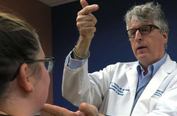
“Patients get a predisposed feeling that their life is doomed. That it’s going to be a complicated life, which isn’t necessarily so,” said Dr. Cary Twyman, a neurologist with Penn State Health. “There are many misconceptions and false information about MS on the internet, so I make sure that each of my patients properly understand what the disease is, how it occurs, and the different courses MS can take.”
The immune system of someone with MS has lost its sense of self. Cells that protect the body from disease can’t detect what is dangerous and what isn’t, so they attack the brain, optic nerve and spinal cord.
Scientists still don’t know what causes MS, but they believe the disease is triggered by an unidentified environmental factor in a person who is genetically predisposed to respond.
The progress and severity of MS and what symptoms it may cause in any one person cannot be predicted. Common ones include numbness or tingling in the face, body or arms and legs; pain; fatigue; walking difficulties; muscle spasms; general weakness; vision problems; and dizziness or vertigo.
Most people with MS find out that they have it between the ages of 20 and 50. At least twice as many women as men are diagnosed.
“MS manifests itself in the prime of these individuals’ lives,” Twyman said. “These people are usually at the ages where raising a family, finding a steady job, and creating different purposes in their lives is a priority. This disorder disrupts that.”
A combination of methods can be used to diagnose MS. These include a careful study of the patient’s medical history, a neurologic exam, and various tests, including MRI, evoked potentials and spinal fluid analysis. Some of these are included in revised guidelines released in 2017 with an aim of providing an earlier diagnosis.
“Over the years, diagnosing MS has become clearer,” Twyman said. “Incidents of MS have doubled since our last census. More than 800,000 people have been diagnosed in the U.S. alone, with approximately 10,000 new cases diagnosed every year, but there has been less misdiagnosis or people going undiagnosed.”
Twyman said finding a treatment to alter the immune system should happen immediately after diagnosis.
“We had an explosion of drugs to treat MS, where we use to have none,” he said. “There are now 15 different drugs for people with MS.”
Because everyone is different, there is no standard way to choose a drug to treat MS. Twyman said finding the right medication for a patient with MS depends on how far along the disease is, what the patient’s tolerance to the risk of the drug is, the cost of the drug, and how closely the patient needs to be monitored on the drug.
In addition to medication, Twyman said caring for a patient with MS has shifted to a team approach.
“It’s no longer a physician-only approach to treat MS,” he said. “It now involves a team that pays attention to an individual’s medical and nonmedical needs to help with their wellness as they live with MS. This team may include nurses, dieticians, social workers and therapists, including the specialties of physical therapy, occupational therapy, speech therapy, and cognitive and behavioral management.”
Twyman said identifying and creating lifestyle habits, such as staying physically fit, reducing stress, and not smoking will help a patient’s quality of life.
“As a comprehensive team, we are able to help our patients with MS look at their life conditions and see what improvements are needed, and then help them make those improvement together,” he said.
It is not true that patients treated with deep brain stimulation (DBS) of the subthalamic nucleus make always more impulsive decisions than others, with major effects on their health and safety. This is what emerges from a study conducted by SISSA in association with the hospitals of Trieste and Udine, Italy. Credit: rawpixel on Unsplash
Promises of food, sums of money or entertaining pastimes: it does not matter what the temptation is, a new study shows that patients suffering from Parkinson’s disease who are treated with Deep Brain Stimulation of the subthalamic nucleus are not more impulsive than others when making decisions about a stimulus that they find particularly appealing. “Deep Brain Stimulation” (DBS) is an effective surgical technique widely used to treat symptoms of Parkinson’s disease. However, the same technique can expose patients to changes in behaviour and in decision-making processes, for example towards food. This alteration could make them adopt risk behaviours. And yet, a study, conducted by a team led by Marilena Aiello and Raffaella Rumiati, Director of Laboratorio Neuroscienze e Società of SISSA, in association with the “Ospedali Riuniti” of Trieste and the “Azienda Ospedaliera Universitaria” Santa Maria della Misericordia of Udine and published on Journal of Neurology, has found that these alterations do not seem to affect all forms of decision. To establish this, the scientists devised and conducted an experiment, which placed the patients in front of a crucial choice: have a small prize immediately or a bigger one, later. The results that emerged from the research add an important element to understanding the disease and the benefits and problems of the DBS technique, opening up interesting clinical and research prospects.
Three groups, three rewards, no difference
“Psychiatric problems such as obsessions or compulsive behaviours, like the tendency to assume unjustified risks in play, to be unable to resist the temptation of food and greater impulsivity, are sometimes observed in patients with Parkinson’s disease treated with DBS, a technique which involves implanting electrodes into the subthalamic nucleus of the brain. It is a consolidated treatment that allows the patients who are treated to reduce the doses of drugs they take, but this can have undesirable side-effects on the cognitive and emotional sphere and on behaviour” explains the scientist Marinella Aiello. To study decisional impulsivity in these patients, which could be what lies behind their risk choices, the research group used what is technically called “delay discounting”: “We put three groups of people—the first composed of Parkinson’s sufferers with DBS, one with Parkinson’s sufferers without DBS, a third composed of healthy people—in front of a choice” explain the scientists. “In a computer exercise they could decide whether to have a small reward immediately, in the form of particularly appealing food, money or facilitations for activities they consider pleasurable. Or the same reward, but in larger quantities later. In these tasks, the choice usually depends on the time that passes between one option and the other: if it is very short, delayed gratification is chosen and vice versa. The principle behind this experiment is the following: the more the impulsive trait is present, the more the first choice will always be preferred over the second. We measured their performance in this task”. No difference emerged between the three groups: “Our study confirms that patients with DBS are no more impulsive in this kind of situation and they do not try to find gratifications more hastily than the others. Moreover, for the first time, we have demonstrated that this does not even depend on the type of reward offered to them”.
The results on patients with eating disorders and weight increase
There is more: “It has been shown that injuries to or stimulations of the subthalamic nucleus increase the motivation to gratify oneself with food. And yet, in our study, impulsive decision making has remained unchanged, even in the people who, after the surgery, had gained weight or had eating problems compared with those who had none of these undesirable effects. And this is very interesting scientifically speaking”. Instead, explains Aiello, “an increase in impulsivity is observed in patients with fewer years from DBS surgery, with higher doses of levodopa—substance used to treat the symptoms of Parkinson’s disease—with higher memory performance. By revealing interesting relationships between the therapeutic treatments and specific behaviours of the patients, our results contribute to shedding light on the clinical results of such an important treatment like DBS for Parkinson’s disease”.
Alzheimer’s-affected brains are riddled with amyloid plaques, protein aggregates consisting mainly of amyloid-β. However, amyloid-β is a fragment produced from a precursor protein whose normal function has remained enigmatic for decades. A team of scientists at VIB and KU Leuven led by professors Joris de Wit and Bart De Strooper has now discovered that this amyloid precursor protein modulates neuronal signal transmission by binding to a specific receptor. Modulating this receptor could potentially help treat Alzheimer’s or other brain diseases. The results are published in Science.
More than 30 years have passed since the amyloid precursor protein was first identified. In the late 1980s, several research teams around the globe traced the protein fragment found in amyloid plaques back to a gene located on chromosome 21. The gene encodes a longer protein that is cleaved into several fragments, one of which ends up in amyloid plaques.
Decades of research have focused on the cleavage process that leads to the formation of the amyloid-β fragment and its subsequent aggregation, in the hope of identifying new therapeutic avenues for Alzheimer’s. Meanwhile, an important question remained unanswered: What does the rest of the amyloid precursor protein actually do?
In search of a binding partner
To answer this question, Dr. Heather Rice, a postdoctoral researcher in the labs of Joris de Wit and Bart De Strooper at the VIB-KU Leuven Center for Brain & Disease Research, set out to identify the nerve cell receptor that interacts with the amyloid precursor protein.
“We knew that the amyloid precursor protein exerts its role through the part of the protein that is released outside of the cell. To understand its function, we needed to look for binding partners located on the cell surface,” explains Rice.
The researchers identified a receptor present at the synapse, the structure where two different neurons connect to pass on signals. “We found that the secreted part of the amyloid precursor protein interacts with a receptor called GABABR1a, and that this in turn suppressed neuronal communication at the synapse,” says Rice.
Modulating signal transmission
“Although mutations in the amyloid precursor protein in familial cases of Alzheimer’s disease all affect the production of amyloid-β, we don’t really know whether other aspects of the protein’s function contribute to Alzheimer’s as well,” says Bart De Strooper. He believes that the new findings add a fresh perspective to previous studies on the amyloid precursor protein and Alzheimer’s disease. “The newly identified role of the amyloid precursor protein may underlie the neuronal network abnormalities we see in mouse models of Alzheimer’s disease and preceding clinical onset in human patients. It is exciting to consider that a therapy targeting this receptor might attenuate these abnormalities in people with Alzheimer’s.”
De Wit adds that the clinical implications may reach much further than just Alzheimer’s: “Interestingly, GABABR signaling has been implicated in a diverse range of neurological and psychiatric disorders, including epilepsy, depression, addiction and schizophrenia. Now that we know how the secreted part of the amyloidprecursor protein modulates neuronal signaling through the GABAB receptor, we could think of new ways to develop drugs that can restore this type of neuronal signaling in other clinical contexts.”
(HealthDay)—Millennials struggling with depression aren’t being helped by their use of Facebook, Instagram or Snapchat, a new study reports.
College students who meet the criteria for major depressive disorder tend to use social media more often and are more heavily addicted to social media, researchers found.
They’re also more likely to use social media in ways that exacerbate or highlight their depression, the study said.
For example, depressed young adults are more likely to compare themselves on social media to people who appear better off than them, said first author Anthony Robinson. He’s a research assistant in the psychology department at Texas State University.
Folks who post to Facebook or Instagram take pains to portray themselves in a flattering light, Robinson said.
“If people are making comparisons based on this inflated image that’s being presented, it can cause feelings of inferiority,” Robinson said.
Depressed young adults were also more likely to be bothered if tagged in an unflattering picture, less likely to post pictures of themselves with other people, and more likely to self-censor what they posted to avoid the judgment of others, results showed.
For the study, Robinson and his colleagues asked 504 undergraduates at Texas State to complete an online survey. The survey assessed their social media use and asked a variety of psychological questions.
Those with symptoms of depression reported behaviors like excessive sleeping, feelings of hopelessness and guilt, or a loss of pleasure in activities they used to enjoy, Robinson said.
About 16 percent of the students met the criteria for major depressive disorder, which Robinson said was an “extremely high” proportion.
Major depressive disorder affects nearly 7 percent of Americans 18 and older in any given year, according to the U.S. National Institute of Mental Health. Young adults 18 to 25 are the age group most likely to have had a major depressive episode in 2016, around 11 percent.
The no-holds-barred, say-anything-without-consequences nature of social media could contribute to people feeling bad about themselves, Robinson said.
“If you’re spending more time on these platforms and you’re being trolled or cyberbullied, it’s going to have a negative effect on your psychological well-being,” Robinson said.
But because this was an observational study, the researchers can’t say in what direction the association between depression and social media works, noted Joseph McGuire, an assistant professor of psychiatry and behavioral sciences at Johns Hopkins Children’s Center in Baltimore. He was not involved with the study.
“Is it that people who tend to be on social media more then start to feel depressed, or is it that the people who are more depressed are more withdrawn and this is their only social contact?” McGuire asked.
“If I spend two hours a day, am I more likely to be a little bit down or depressed compared with someone who is only on 20 minutes a day? Or do I never go out because I’m depressed, but I still want that social contact, so I log on to social media?” he continued.
Another new study backs up these findings. Researchers found that people are more aware of their own physical ailments if they tend to use Facebook a lot and frequently compare themselves to people apparently better off than themselves, according to findings in the January issue of the journal Heliyon.
College students who are feeling depressed should assess their own social media use, and either cut back or try to change their online behaviors if they are using the technology in ways linked to depression, Robinson said.
It wouldn’t hurt at all if they also sought counseling, McGuire said.
“There’s a lot of counseling centers available at academic centers. There’s that support there, and it’s intended to help kids who are struggling,” McGuire said. “Reaching out to a professional, even if you don’t think you have a problem, then you can at least start to have that dialogue.”
The new study was published Jan. 9 in the Journal of Applied Biobehavioral Research.
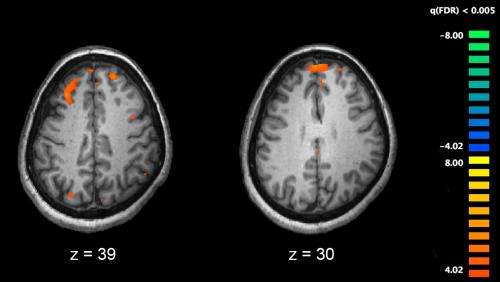
Functional magnetic resonance imaging (fMRI) and other brain imaging technologies allow for the study of differences in brain activity in people diagnosed with schizophrenia. The image shows two levels of the brain, with areas that were more active in healthy controls than in schizophrenia patients shown in orange, during an fMRI study of working memory. Credit: Kim J, Matthews NL, Park S./PLoS One.
Despite extensive research efforts, schizophrenia remains one of the least understood brain disorders. One promising area of research is in receptors on the surfaces of brain cells that help sense growth factors. But there’s been a problem: in previous schizophrenia studies, researchers have genetically manipulated brain cell receptors in very young mice. Schizophrenia usually affects adults.
In a recent issue of the Proceedings of the National Academy of Sciences, Lin Mei, MD, Ph.D., asked, does all the tinkering in young mice hamper their brain development, causing schizophrenia-like symptoms? Or, do their brain cells develop normally, but in adulthood struggle to communicate? Researchers need to know whether to focus their efforts on brain cell development or communication, or both, because the answer to these questions implies different therapeutic approaches.
In the new study, Mei, professor and chair of neurosciences at Case Western Reserve University School of Medicine, led an international team of neuroscientists. The team included Mei’s long-time collaborator, Wen-Cheng Xiong, Ph.D., professor of neurosciences, and first authors Hongsheng Wang and Wenbing Chen, graduate students, all of CWRU. Additional collaborators included researchers at Nanchang University and Guangzhou Medical University in China, and neuroscientists from the Medical College of Georgia at Augusta University.
Together, the researchers studied a brain cell receptor—ErbB4—whose level is altered in adults with schizophrenia. ErbB4 helps maintain an inhibitory neurotransmitter in the brain—GABA—that prevents brain cells from overreacting and keeps fear and anxiety in check. The researchers have shown previously that ErbB4 mutations change signals inside brain cells that lead to schizophrenic symptoms in mice.
“When ErbB4 is mutated early on in mice, it impairs brain circuit wiring. It also impairs GABA transmission in adult animals, causing schizophrenic symptoms,” said Mei. “But previous models are unable to distinguish whether deficits are from abnormal development in young mice brains, or abnormal transmission developed later on.” Mei’s new study shows schizophrenic symptoms come from deficits in how brain cells communicate during adulthood, regardless of whether or not they fully developed.
To find their answers, Mei’s team genetically engineered two new mouse models of schizophrenia. In the first, the researchers treated mice with a chemical that switches “off” the gene encoding ErbB4. “Using inducible knock-out mice, we depleted ErbB4 only in adult animals, and showed that this impairs behavior,” said Mei. In mice missing ErbB4 only in adulthood, brain cell development and appearance were normal, but symptoms persisted. The experiment suggested schizophrenic symptoms in adult mice were unrelated to abnormal brain cell development.
In the second mouse model, the receptor was missing in mice from the beginning, hampering brain cell development. The researchers used the same genetic switch to turn ErbB4 “on” in adulthood—in essence, recovering it. “In recovery knock-out mice, ErbB4 is missing during development and thus the mice have crippled brain circuits. Yet, when ErbB4 is restored on a malformed circuit, mice scored better in behavioral tests,” said Mei. Even with underdeveloped brain cells, schizophrenic symptoms could be alleviated simply by adding ErbB4.
Mei’s team found restoring ErbB4 receptors reduced hyperactivity, and normalized fear responses in adult mice. “ErbB4 is a risk factor for schizophrenia,” said Mei. “This study shows correcting ErbB4 signaling could be therapeutic in relevant patients.”
The results in the two mouse models confirm that ErbB4 is critical to how brain cells communicate during adulthood. The nuanced distinction could lead to new therapeutics designed to improve brain cell signaling associated with the ErbB4 receptor. In particular, therapeutics that improve how GABA neurotransmitters regulate brain cell activity.
“Restoring ErbB4 could be beneficial to patients—even those with malformed brain circuitry,” said Mei. “We are now looking into how restoring ErbB4 improves neurotransmitter signaling inside brain cells, including those relevant to other psychiatric disorders, such as attention deficit hyperactivity disorder and major depression.”
The kicks a mother feels from her unborn child may allow the baby to ‘map’ their own body and enable them to eventually explore their surroundings, suggests new research led by UCL in collaboration with UCLH.
For the study, published today in Scientific Reports, researchers measured brainwaves produced when newborn babies kick their limbs during rapid eye movement (REM) sleep, finding that fast brainwaves—a brainwave pattern typically seen in neonates—fire in the corresponding hemisphere.
For example, the movement of a baby’s right hand causes brainwaves to fire immediately afterwards in the part of the left brain hemisphere that processes touch for the right hand. The size of these brainwaves is largest in premature babies, who at that age would usually still be in the womb.
The findings suggest that foetal kicks in the late stages of pregnancy—the third trimester—help to grow areas of the brain that deal with sensory input, and are how the baby develops a sense of their own body. The fast brainwaves evoked by the movement disappear by the time babies are a few weeks old.
“Spontaneous movement and consequent feedback from the environment during the early developmental period are known to be necessary for proper brain mapping in animals such as rats. Here we showed that this may be true in humans too,” explained study author Dr. Lorenzo Fabrizi (UCL Neuroscience, Physiology & Pharmacology).
This video shows how fast brainwaves are produced when a newborn moves a limb. Credit: Kimberley Whitehead, Lorezo Fabrizi, Judith Meek
Kimberley Whitehead (UCL Neuroscience, Physiology & Pharmacology) said: “We think the findings have implications for providing the optimal hospital environment for infants born early, so that they receive appropriate sensory input. For example, it is already routine for infants to be ‘nested’ in their cots—this allows them to ‘feel’ a surface when their limbs kick, as if they were still inside the womb.
“As the movements we observed occur during sleep, our results support other studies which indicate that sleep should be protected in newborns, for example by minimising the disturbance associated with necessary medical procedures.”
The babies’ brainwaves were measured using electroencephalography (EEG), and were recorded continuously during sleep. Active sleep was identified behaviourally according to cot side observation of rapid eye movements, largely irregular breathing and frequent, isolated limb movements.
This video shows how fast brainwaves are produced when the newborn moves a limb. Credit: Kimberley Whitehead, Lorenzo Fabrizi, Judith Meek
A total of 19 newborns aged two days on average took part in the study, and they were between 31 and 42 weeks corrected gestational age when studied. Corrected gestational age takes into account their age if they were still in the womb; a baby born at 35 weeks and being one week old would have a corrected gestational age of 36 weeks.
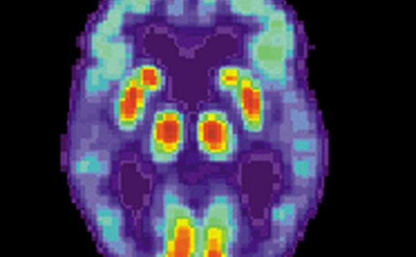
PET scan of a human brain with Alzheimer’s disease. Credit: public domain
A major new study on Alzheimer’s disease provides previously unknown evidence of how the brain-robbing illness may originate.
Moreover, it proposes that certain HIV drugs called reverse transcriptase inhibitors could immediately be repurposed for Alzheimer’s patients.
Led by scientists from Sanford Burnham Prebys Medical Discovery Institute in San Diego, the study finds that, as long suspected, Alzheimer’s is a genetic disease. But in nearly all cases, it’s not inherited. Rather, it arises during a patient’s lifetime by genetic rearrangements in neurons. Sequences of DNA are copied, altered and inserted back into the genome.
The genetic rearranging isn’t random mutation, but a process that recombines DNA into different patterns. This reshuffling creates a mosaic of slightly differing cells. The immune system uses a similar process to make antibodies, but nothing like it has been seen in the human brain.
Reverse transcriptase inhibitors might also ward off Alzheimer’s in those with Down syndrome, who develop Alzheimer’s as they age, the study said.
The study was published Wednesday in the journal Nature.
Confirmation of the findings is required, said Dr. Jerold Chun, the lead author. But Chun says testing with the HIV drugs should begin immediately. Even a low degree of effectiveness would be better than what is now available.
The study combines single and multiple-cell analytical methods to examine 13 donated human brains, some normal, some with Alzheimer’s. Its findings jibe with epidemiological data from elderly HIV patients. They have been treated with reverse transcriptase inhibitors for decades, and almost never get Alzheimer’s.
The first documented case of Alzheimer’s in an HIV-positive individual was reported in 2016.
Cautious praise for the study came from Dr. Paul Aisen, a longtime Alzheimer’s researcher who specializes in clinical trials. Aisen heads the University of Southern California Alzheimer’s Therapeutic Research Institute in San Diego.
“The authors carefully demonstrate that there are extensive modifications to genetic material in the Alzheimer’s disease brain,” Aisen said by email.
“These are changes that occur with aging, rather than inherited genetic characteristics. While this is an intriguing idea, the actual contribution of this age-related genetic change remains uncertain.”
Fred “Rusty” Gage, president of the Salk Institute and a noted brain expert, said the study’s results back up the claim that DNA sequences are copied and inserted back into the neuronal genome.
“These results are quite striking and could have implications for Alzheimer’s disease diagnosis and progression,” Gage said by email.
About 5.7 million Americans today have been diagnosed with Alzheimer’s disease, according to the Alzheimer’s Association. That number is expected to double by 2060, according to the Centers for Disease Control and Prevention.
In recent years, Alzheimer’s researchers have changed their view of the disease. They now say Alzheimer’s begins decades before symptoms appear. Eventually, the damage eating away at the brain becomes severe enough to affect cognition and memory.
So increasingly, researchers are looking for the earliest possible signs that Alzheimer’s is developing, before mental functions are affected.
The study traces the ultimate cause to the genetic rearrangements, so blocking this reshuffling should block Alzheimer’s.
The reshuffling can be likened to a copy-and-paste function in affected neurons. But instead of making an exact copy, the process scrambles DNA segments, then reinserts them back into the neuron’s genome.
Normal brains also show genetic variation in individual cells. Research suggests this condition is a normal part of brain development. Instead of having billions of identical neurons, each may vary slightly in a way that helps the brain work, Chun said.
This process goes wrong in producing the Alzheimer’s-causing variations, derived from a gene called APP. Certain variants of this gene are strongly linked with Alzheimer’s.
Because genes produce proteins, these rearrangements of the APP gene likely produce variations of toxic brain proteins called beta amyloid, known to be involved in Alzheimer’s.
Some of these genetic variants are found in a very rare form of Alzheimer’s that is directly inherited. Virtually all people with these variations come down with Alzheimer’s.
But this “familial” form constitutes only a few percent of all Alzheimer’s cases. The vast majority of “sporadic” Alzheimer’s cases shows genetic tendencies, but fall far short of a perfect correlation.
The study provides an explanation for sporadic Alzheimer’s: Because these genetic changes only occur in the brain, they don’t show up when a person’s genome is sequenced.
In addition, it may explain the failures of amyloid-based therapy. Billions of dollars have been spent developing drugs according to what is known as the “amyloid hypothesis,” with virtually nothing to show for it.
Chun said this may be because the amyloid drugs are aimed at a single molecular target, and there’s molecular diversity in amyloid.
“What our data strongly supports is that there could be many, many other targets that would have been missed by these single molecularly targeted therapeutics,” Chun said.
This means the amyloid hypothesis is essentially correct, but doesn’t go deep enough, Chun said.
In an accompanying perspective article, two University of California, San Diego Alzheimer’s researchers said the study was also important because it provided the “surprising existence” in the brain of what is called “somatic gene recombination.”
“This phenomenon, which has previously been reported only in antibody generation in immune cells, increases the diversity of proteins encoded by a given gene through DNA-shuffling mechanisms, wrote Guoliang Chai and Joseph G. Gleeson.
“The study hints at a previously unanticipated mechanism in the development of Alzheimer’s, and expands our understanding of the genesis of brain mosaicism,” they wrote. “But whether accumulation of (the recombined DNA) in neurons is a cause of or is caused by Alzheimer’s disease remains to be proved.”
Aisen cautioned that some studies indicate that Alzheimer’s is more likely linked to defects in removing amyloid, not their manufacture.
“So while there could be therapeutic implications of these new findings, additional clarification of this mechanism is needed,” Aisen said.
Doctors could use the HIV drugs on Alzheimer’s patients as an “off-label” use, Chun said. But that would require careful ethical consideration.
On the positive side, there’s no treatment now that affects the underlying neurodegeneration Alzheimer’s brings, and the safety profile of the HIV drugs is well-known.
“Let’s say this only works 25 percent of the time, that’s still 1.5 million patients in the United States, not to speak of their families,” he said.
And even a modest effect of preserving cognition for a few years would mean a great deal to those families, he said.
However, all drugs have risks, and Alzheimer’s patients tend to be elderly and may have other conditions.
Such off-label use also doesn’t provide a scientific basis for officially approving the drugs for Alzheimer’s. That’s the job of a properly designed clinical trial.
Designing such a trial will be complicated, Chun said. There’s the need to accurately find the patients who will likely benefit. Brain scans for amyloid deposits, which are now available, might help. But as for now, actual detection of the pathological brain mosaicism in living people isn’t possible.
The APP-linked variations spread with the help of an enzyme called reverse transcriptase that HIV uses to replicate, the study says.
With the help of reverse transcriptase, HIV copies its genome into the genome of infected cells. Since it becomes part of the cell’s DNA, HIV is very hard for the immune system to reach. Reverse transcriptase inhibitors, a regular part of HIV therapy, block this process.
Reverse transcriptase is also naturally found in the human body, and presumably plays an important function.
“Evolutionary biologists that have looked at the human genome, have estimated that nearly half of our genome was created by reverse transcription,” he said.
Whether HIV’s reverse transcriptase is identical to the human version isn’t known, Chun said.
Research funding was provided by The Shaffer Family Foundation; The Bruce Ford & Anne Smith Bundy Foundation; Sanford Burnham Prebys; the National Institutes of Health; and the government of Taiwan.
Highlighted areas show brain regions where researchers saw reductions in brain volume in girls who self-injure. Credit: The Ohio State University
The brains of teenage girls who engage in serious forms of self-harm, including cutting, show features similar to those seen in adults with borderline personality disorder, a severe and hard-to-treat mental illness, a new study has found.
Reduced brain volumes seen in these girls confirms biological—and not just behavioral—changes and should prompt additional efforts to prevent and treat self-inflicted injury, a known risk factor for suicide, said study lead author Theodore Beauchaine, a professor of psychology at The Ohio State University.
This research is the first to highlight physical changes in the brain in teenage girls who harm themselves.
The findings are especially important given recent increases in self-harm in the U.S., which now affects as many as 20 percent of adolescents and is being seen earlier in childhood, Beauchaine said.
“Girls are initiating self-injury at younger and younger ages, many before age 10,” he said.
Cutting and other forms of self-harm often precede suicide, which increased among 10- to 14-year-old girls by 300 percent from 1999 to 2014, according to data from the Centers for Disease Control and Prevention. During that same time, there was a 53 percent increase in suicide in older teen girls and young women. Self-injury also has been linked to later diagnosis of depression and borderline personality disorder.
In adults with borderline personality disorder, structural and functional abnormalities are well-documented in several areas of the brain that help regulate emotions.
But until this research, nobody had looked at the brains of adolescents who engage in self-harm to see if there are similar changes.
The new study, which appears in the journal Development and Psychopathology, included 20 teenage girls with a history of severe self-injury and 20 girls with no history of self-harm. Each girl underwent magnetic resonance imaging of her brain. When the researchers compared overall brain volumes of the 20 self-injuring girls with those in the control group, they found clear decreases in volume in parts of the brain called the insular cortex and inferior frontal gyrus.
These regions, which are next to one another, are two of several areas where brain volumes are smaller in adults with borderline personality disorder, or BPD, which, like cutting and other forms of self-harm, is more common among females. Brain volume losses are also well-documented in people who’ve undergone abuse, neglect and trauma, Beauchaine said.
The study also found a correlation between brain volume and the girls’ self-reported levels of emotion dysregulation, which were gathered during interviews prior to the brain scans.
Beauchaine said this study does not mean that all girls who harm themselves will go on to develop BPD—but it does highlight a clear need to do a better job with prevention and early intervention.
“These girls are at high risk for eventual suicide. Self-injury is the strongest predictor of suicide outside of previous suicide attempts,” Beauchaine said. “But there’s most likely an opportunity here to prevent that. We know that these brain regions are really sensitive to outside factors, both positive and negative, and that they continue to develop all the way into the mid-20s,” he said.
Adolescents who self-injure are more anxious, more depressed and more hostile than their peers who are also referred to mental health experts, previous studies have shown. This new brain-volume evidence bolsters the argument that self-injury should be viewed as a potential sign of serious, life-threatening illness, Beauchaine said, adding that there are no major prevention projects currently aimed at pre-adolescent girls in the United States. Instead, most current interventions start in adolescence when risk for self-harm is greatest.
“A lot of people react to girls who cut by saying, ‘She’s just doing it for attention, she should just knock it off,’ but we need to take this seriously and focus on prevention. It’s far easier to prevent a problem than to reverse it,” he said.
He said it’s important to recognize that the research does not establish whether the decreased brain volume seen in the study preceded the self-harm, or emerged after the girls began to injure themselves.
Further studies looking at brain changes are needed to help researchers better understand the relationship between structural differences and self-harm and how those might correspond to BPD and other mental disorders down the road, Beauchaine said.
“If we can learn more about how adults with psychiatric disorders got there, we are in a much better position to take care of people with these illnesses, or even stop them from happening in the first place,” he said.
A previously published study in these same girls applied functional MRI during a task in which they could receive monetary rewards. The researchers saw diminished brain responses to reward in those girls with a history of self-harm—results that looked similar to previous studies of adults with mood disorders and borderline personality disorder.
“Self-injury is a phenomenon that’s increasing, and that’s less common outside of the United States. It’s saying something about our culture that this is happening, and we should do whatever we can to look for ways to prevent it,” Beauchaine said.
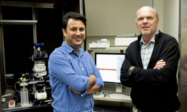
A research team led by Harald Sontheimer (right), a professor at the Virginia Tech Carilion Research Institute, has discovered mysterious brain structures called perineuronal nets help modulate electrical impulse activity in the brain. The team, which include first author and postdoctoral fellow Bhanu Tewari (left), may be able to target the nets to treat some cases of acquired epilepsy. Credit: Virginia Tech
Scientists at the Virginia Tech Carilion Research Institute have solved a 125-year-old mystery of the brain, and, in the process, uncovered a potential treatment for acquired epilepsy.
Since 1893, scientists have known about enigmatic structures called perineuronal nets wrapped around neurons, but the function of the nets remained elusive.
Now, a research team led by Harald Sontheimer, the director of the VTCRI Center for Glial Biology in Health, Disease, and Cancer and the executive director of the School of Neuroscience, part of the Virginia Tech College of Science, has determined the nets modulate electrical impulses in the brain. What’s more, brain seizures can occur if the nets are dissolved.
The discovery, published today in Nature Communications, has implications in various forms of acquired epilepsy, a type of seizure disorder that results from brain lesions caused by trauma, infection, or tumors in the brain.
“We started by investigating tumor-associated epilepsy, and we accidentally learned something else important about how the brain works in disease and in health,” Sontheimer said.
The researchers initially made their finding in a mouse model of epilepsy caused by the deadly brain cancer known as glioblastoma, the first symptom of which is often a seizure.
Glioblastoma is the only cancer whose growth is restricted by space. Since the skull blocks the cancer from expanding outward, the tumor produces an excitatory chemical neurotransmitter called glutamate in excessive amounts that kills neighboring healthy cells to make room to grow.
The researchers saw that glutamate targeted brain cells producing a different chemical neurotransmitter called “GABA,” that usually calms neurons by inhibiting them from firing electrical impulses once the messages are relayed. Without GABA, the brain becomes too excited and can seize.
In addition to glutamate, the tumor also secretes an enzyme aimed at destroying the surrounding extracellular matrix, a gel-like substance that holds brain cells in place. Glioblastomas are highly malignant and notoriously invasive—the enzyme is the knife that cuts the cancer’s tethers and lets it migrate freely.
“Unexpectedly, we also saw the enzyme attacking the perineuronal nets,” Sontheimer said, noting that the nets are primarily found wrapped around the GABA-secreting inhibitory neurons, which help prevent seizures. “It was a surprise to see this bystander effect of seizure activity once the neurons were stripped of their nets.”
Italian neurobiologist Camillo Golgi was the first to identify perineuronal nets in 1893, but he misunderstood their function. Golgi called the nets “corsets,” and said they most likely impeded messaging between neurons.
To the contrary, Sontheimer found the nets enabled messaging. The neurons covered by perineuronal nets have a reduced membrane capacitance or ability to store electrical charge, meaning that they can fire an impulse and reload up to twice as fast as non-netted neurons.
When the inhibitory neurons suddenly lose their perineuronal nets, the results can be catastrophic. The researchers applied the enzyme to brains without tumors and saw that on its own, the enzymatic degradation of the perineuronal nets was enough to induce seizures—even when the neurons were left intact.
“Without the perineuronal nets, inhibitory neurons would fire too slowly and therefore inhibition becomes too little, too late, and a seizure will occur—even in otherwise healthy brains,” Sontheimer said, noting that the enzyme can devour a perineuronal net in less than 30 minutes. “No one thought that these structures would have such a profound effect on how normal processes operate.”
Now, the researchers are studying how perineuronal nets might play a role in other forms of acquired epilepsy, which can result from head injury or brain infection. Such elucidation could lead researchers to discover potential pharmacological solutions.
“Importantly, the finding that tumor-induced disruption in perineuronal nets contributes to imbalanced inhibitory neurotransmission suggests a new target for therapeutic intervention to control tumor-associated seizures,” said H. Steve White, a professor and chair of the Department of Pharmacy in the University of Washington’s School of Pharmacy in Seattle, Washington.
White, a renowned expert in epilepsy and anticonvulsant drug development research, was not involved in Sontheimer’s study.
“Although additional studies are needed, it is likely that the findings reported by Dr. Sontheimer and his team are applicable to other forms of acquired epilepsy where brain injury is associated with a heightened inflammatory response,” White said, noting that the implications for treatment and prevention of epilepsy are particularly striking since current therapies are aimed at controlling seizures. “While controlling the symptoms of the disease is significant, the results of this study suggest a possible path toward modifying the development and progression of epilepsy, which would lessen the overall burden to the patient.”
More than 50 million people worldwide have epilepsy, according to the World Health Organization. About a third of those individuals don’t respond to current anti-epileptic treatments.
“If we confirm our hypothesis that digested perineuronal nets are responsible for other forms of acquired epilepsy, then a potential treatment could be an enzyme inhibitor,” Sontheimer said.
He noted that one such an inhibitor is already approved by the FDA for other uses but he also cautioned that there’s a significant amount of research to conduct before their hypothesis is confirmed.
“We need new approaches to treat epilepsy. I think this could be an effective way to control seizures,” Sontheimer said. “And we solved a 125-year-old neuroscience mystery! This is what basic science is all about—keeping an open and observant mind to answer questions old and new.”
Genetics may predispose some people to both Alzheimer’s disease and high levels of blood lipids such as cholesterol, a common feature of cardiovascular disease, according to a new study by an international team of researchers led by scientists at UC San Francisco and Washington University School of Medicine in St. Louis.
The research analyzed genome-wide data from over 1.5 million individuals, making it one of the largest-ever studies of Alzheimer’s genetics. The authors hope the findings will lead to improved early diagnosis and potentially new preventative strategies for Alzheimer’s disease, which currently affects 5.7 million people in the U.S. and has no cure.
Mounting clinical and epidemiological evidence has pointed to a link between heart disease and Alzheimer’s disease, but a biological relationship between the two conditions has remained controversial. Many patients diagnosed with Alzheimer’s disease also show signs of cardiovascular disease, and postmortem studies reveal that the brains of many Alzheimer’s patients show signs of vascular disease, which some scientists speculate could drive the onset of dementia. These observations led to hopes of preventing Alzheimer’s by treating cardiovascular symptoms, but initial genetic studies and failed clinical trials of cardiovascular drugs called statins in Alzheimer’s disease have cast doubt on this possibility.
The new study, published November 9, 2018 in Acta Neuropathologica, shows that Alzheimer’s and cardiovascular disease do share common genetics in some individuals, raising new questions about whether this shared biology could be targeted to slow down or prevent both diseases.
“These results imply that irrespective of what causes what, cardiovascular and Alzheimer’s pathology co-occur because they are linked genetically. That is, if you carry this handful of gene variants you may be at risk for not only heart disease but also Alzheimer’s,” said UCSF neurodegeneration researcher Rahul Desikan, MD, Ph.D., a clinician-scientist whose lab is known for developing a so-called polygenic hazard score for Alzheimer’s that predicts the age at which an individual is likely to begin experiencing symptoms of dementia based on their genetic background.
To identify genetic variants that confer risk of both cardiovascular disease and Alzheimer’s disease, the researchers used statistical techniques pioneered by Desikan’s lab in collaboration with Ole Andreassen, MD, Ph.D., at the University of Oslo, and Anders Dale, Ph.D., at UC San Diego, that allowed them to combine multiple large-scale genome-wide association studies (GWAS)—a type of genetic study that makes statistical links between various disease states and widely shared variations in the genetic code.
Ultimately Desikan’s team was able to analyze the combined impact of such genetic markers on both cardiovascular disease risk—based on five GWAS studies of more than one million individuals—and on Alzheimer’s risk—based on three GWAS studies of nearly 30,000 Alzheimer’s patients and more than 50,000 age-matched controls.
“Our approach works by combining and leveraging these massive GWAS studies to make discoveries that would otherwise be invisible,” Desikan said. “We are essentially borrowing statistical power.”
This analysis enabled the researchers to identify 90 spots in the genome where specific DNA variants increased patients’ combined chance of developing both Alzheimer’s disease and heightened blood levels of lipid molecules, including HDL and LDL cholesterol and triglycerides, which are common risk factors for cardiovascular disease.
The researchers confirmed that six of these 90 regions had very strong “genome-wide significant” effects on Alzheimer’s and heightened blood lipid levels, including several within genes that had never before been linked to dementia risk. These included several sites within the CELF1/MTCH2/SPI1 region on chromosome 11, which had previously been linked to immune system biology.
In contrast, though patients diagnosed with Alzheimer’s also often exhibit other cardiovascular risk factors, such as unhealthy levels of belly fat, type 2 diabetes, and chest pain or other symptoms of coronary artery disease, the authors could find no clear overlapping genetics between Alzheimer’s disease and these risk factors.
“These results suggest that Alzheimer’s and cardiovascular disease could both be influenced by genetic defects that impair the body’s ability to processes lipids properly,” said study first author Iris Broce-Diaz, Ph.D., a postdoctoral researcher in Desikan’s lab. “But they also suggest that the link between Alzheimer’s and other cardiovascular risk factors are not likely due to common genetics, though they could be linked by diet or other lifestyle factors.”
“This is exciting because we know that levels of cholesterol and other lipids in the blood are highly modifiable through changes in diet or with drugs,” Broce added. “This raises the possibility that we might be able to help delay or even prevent the onset of Alzheimer’s in these patients, though we’ll need more data before we can say that for sure.”
Desikan’s team then examined whether currently healthy individuals with a family history of Alzheimer’s disease were more likely to carry these newfound genetic variants, a relatively new method for studying early Alzheimer’s risk factors called “Alzheimer’s-by-proxy.” Taking advantage of a large British research cohort called UK Biobank, the researchers found these variants were much more likely to be present in the genomes of 50,000 individuals who had one or more parents diagnosed with Alzheimer’s disease than in nearly 330,000 individuals without a family history of the disease.
“It’s remarkable that among these healthy adults with a family history of Alzheimer’s, a subset of cardiovascular risk genes themselves appear to strongly influence risk of eventually developing Alzheimer’s,” Desikan said. “This is exactly the population we should study to see if reducing cardiovascular disease risk through lifestyle or medication can delay or prevent the onset of dementia later in life.”
The researchers further confirmed their findings in a collaboration with Celeste M. Karch, Ph.D., an Alzheimer’s expert at Washington University School of Medicine in St. Louis and co-corresponding author on the new study. Karch’s team showed that the novel genes most closely tied to Alzheimer’s/cardiovascular risk in the new study were differently expressed in the brains of Alzheimer’s patients compared to control brains.
“This study emphasizes that we should be thinking about Alzheimer’s disease holistically,” Karch said. “There’s a lot to learn about how the genes that are driving Alzheimer’s disease risk also are driving changes throughout the body.”
“This is also an exciting opportunity because we already know a great deal about these cardiovascular traits and how to target them,” she added.
Desikan and Broce-Diaz are now integrating these findings into the lab’s previously developed polygenic risk score, which will allow clinicians to calculate, based on a patient’s genome, their combined risk for cardiovascular disease and Alzheimer’s. The researchers hope that their work will help launch a precision medicine approach to testing whether controlling blood lipid levels in individuals with the newly discovered risk factors, either through diet or using established drugs such as statins, might delay or prevent the onset of Alzheimer’s disease.
“That’s really the goal,” said Broce-Diaz. “If we can identify the subset of individuals whose cardiovascular and brain health is linked genetically, we think there’s a possibility that reducing their blood lipid levels could help reduce their risk of developing dementia later in life. Such treatments haven’t worked in clinical trials before, but this could be because we haven’t had a good way of selecting who is most likely to benefit based on their genetics.”