http://www.youtube.com/watch?v=X6LqL0WsE80


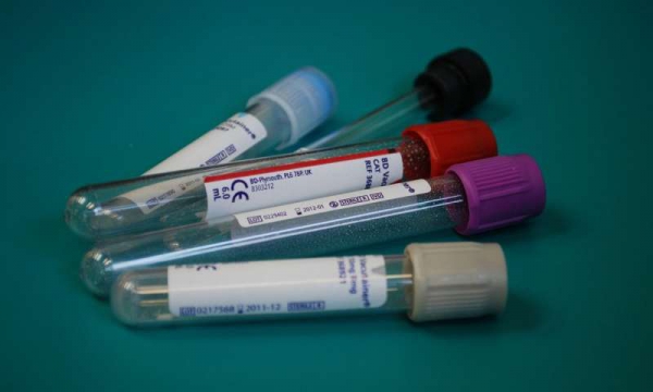
A cancer blood test based on DNA fragment size

Credit: CC0 Public Domain
A team of researchers from the U.K., Denmark, Poland, the Netherlands and Switzerland has developed a new way to test for cancer—by looking at the size of tumor DNA fragments circulating in the bloodstream. In their paper published in the journal Science Translational Medicine, the group describes their technique and how well it worked when tested on patients with different kinds of cancer. Ellen Heitzer and Michael Speicher, with Medical University of Graz, offer a Focus piece on the work done by the team in the same journal issue.
As Heitzer and Speicher note, human blood contains many small DNA fragments, most of them from white blood cells. But there are instances where other types are introduced, for instance, DNA from a fetus in the blood of pregnant women, or bits of DNA that break loose from cancerous tumors. In this new effort, the researchers discovered that DNA from tumors can be differentiated from other types of DNA in the blood by size—such fragments tend to be smaller than DNA fragments from other sources.
To take advantage of this finding, the researchers carefully studied DNA fragments in the blood from several types of cancer and found they could be classified by size. They then created a new metric of measurement for them based on size. They called it trimmed median absolute deviation from copy number neutrality (t-MAD). The team then tested their approach by introducing DNA fragments into a blood sample and then used a benchtop fluidic device to focus on the DNA fragments, which they assigned a t-MAD rating. Pleased with their findings, they further tested their approach by conducting similar tests with blood from actual cancer patients.
The researchers report that their testing showed the technique to be very reliable—it offered positive results for 94 percent of breast, bowel, ovary, skin and bile duct cancers with a false positive rate of just 2.5 percent. It also proved to be 65 percent accurate in detecting pancreas, kidney and brain cancers.
Heitzer and Speicher point out that there are still some questions regarding the technique, however. Chief among them is whether testing will be as accurate when used in a large multicenter clinical setting.
![]()

Journal reference:
Science Translational Medicine
![]()
![]()
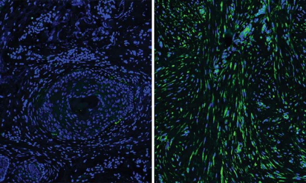
Cells beneath the skin explain differences in healing

A microscopic comparison between normal skin (left) and scarred skin. Credit: Yale University
Differences in the cells that give skin its resilience and strength during wound repair may explain why individuals heal differently, according to a new Yale study published Nov. 23 in the journal Science.
Fibroblasts, the cells that form the protein structure beneath the surface of the skin, were once thought to be fairly uniform in their function. However, the new study found that subsets of fibroblasts may explain why skin regeneration is less robust in older people and how certain types of scars form.
“These subsets of cells may explain different healing potentials in different people,” said senior author Valerie Horsley, associate professor of molecular, cellular, and developmental biology.
Horsley and first author Brett Shook studied the genetics of fibroblasts and looked at their effects in mice and humans and found multiple differences in how these cells respond to injury and changes that occur during aging. For instance, after injury, a subset of fibroblasts that normally produce adipocytes, or fat cells, start forming scar tissue to repair the skin tissue. Interestingly, say the researchers, these cells are reduced in human skin compared to mouse skin, which may explain why scars are more prevalent in people than mice.
The study also identified the essential role of immune cells called macrophages in not only fighting infection but also healing wounds and scar formation. Macrophages that appear at the peak of tissue regeneration can selectively signal to a subset of fibroblasts. Human scar tissue contains more macrophages and fibroblast subsets and both populations are reduced in poorly healing wounds from aged mice.
“By identifying functionally distinct populations of skin cells and revealing the signals that control their behavior, we hope to develop new targeted therapies for the treatment of defective wound healing and scar formation,” Shook said.
![]()
Journal reference:
Science
![]()
![]()
Provided by:
Yale University
![]()
![]()
Breast cancers enhance their growth by recruiting cells from bone marrow

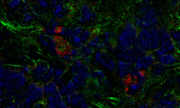
A mouse breast tumor contains bone marrow-derived fibroblasts (red) as well as other cancer-associated fibroblasts (green). Credit: Raz et al., 2018
Researchers in Israel have discovered that breast tumors can boost their growth by recruiting stromal cells originally formed in the bone marrow. The study, which will be published November 23 in the Journal of Experimental Medicine, reveals that the recruitment of bone marrow-derived fibroblasts lowers the odds of surviving breast cancer, but suggests that targeting these cells could be an effective way of treating the disease.
Within solid tumors, cancer cells are surrounded by other cell types that, though not cancerous themselves, boost tumor growth and metastasis. Breast tumors, for example, contain large numbers of fibroblast cells that promote cancer cell proliferation, inflammation, and the formation of new blood vessels to supply the growing tumor with nutrients and oxygen. Many of these cancer-associated fibroblasts are derived from the neighboring breast tissue, but others seem to come from elsewhere in the body.
Neta Erez and colleagues at the Sackler School of Medicine, Tel Aviv University, discovered that, in mice with breast cancer, a significant number of cancer-associated fibroblasts are derived from bone marrow cells called mesenchymal stromal cells (MSCs). The researchers found that breast tumors can recruit MSCs from the bone marrow and cause them to develop into fibroblasts.
These bone marrow-derived fibroblasts are different from other cancer-associated fibroblasts. They lack, for example, a key cell signaling protein called PDGFRα. But bone marrow-derived fibroblasts are particularly effective at stimulating the formation of new blood vessels because they produce large amounts of a protein called clusterin. Tumors containing bone marrow-derived fibroblasts were therefore more vascularized and grew faster than tumors that only contained breast-derived fibroblasts.
Erez and colleagues found that human breast cancers also contain fibroblasts lacking PDGFRα, suggesting that human tumors may also recruit bone marrow-derived cells. Moreover, tumors containing lower levels of PDGFRα tended to be more deadly, suggesting that the recruitment of bone marrow-derived fibroblasts is a crucial step in breast cancer progression.
“Our study shows that the recruitment of bone marrow-derived fibroblasts is important for promoting tumor growth, likely by enhancing blood vessel formation,” Erez says. “Understanding the function of these cancer-associated fibroblasts could form the basis of developing novel therapeutic manipulations that co-target bone marrow-derived fibroblasts as well as the cancer cells themselves.”
![]()
Journal reference:
Journal of Experimental Medicine
![]()
![]()
Provided by:
Rockefeller University Press
![]()
![]()

Study finds a relationship between cold weather and drinking alcohol

Credit: CC0 Public Domain
Now that our days are getting colder and shorter, a new study from the University of Pittsburgh gives us reason to think about the odd relationship between alcohol and weather.
It found that throughout the world and in the United States, drinking levels and liver disease correlated with climate and sunlight. Drinking and disease rose as average temperatures and hours of sunlight fell.
The study, which was published in the journal Hepatology, has public health implications at a time when deaths from cirrhosis of the liver have been rising, particularly among 25- to 34-year-olds. Ramon Bataller, senior author and chief of hepatology at University of Pittsburgh Medical Center, said knowing that heavy drinking is more common in colder climes could help officials who want to reduce damage from alcohol to direct resources toward regions at the highest risk. He also suggested that someone with a family history of alcoholism who has a choice between jobs in, say, Missouri and Minnesota would do well to pick the warmer state.
The World Health Organization estimates that almost 6 percent of deaths throughout the world can be attributed to alcohol misuse.
Bataller said people in colder climates may drink more because alcohol tends to make them feel warmer. On the other hand, people in hot places are more likely to feel uncomfortable or light-headed when they drink. For many people, darkness can also exacerbate depression, which is associated with drinking, though alcohol is a depressant. Snowy climates might also increase isolation, which can make depression worse.
While people in frigid places like Russia are known for heavy drinking, Bataller said the connection between drinking and climate had not been studied systematically before. A team led by Meritxell Ventura-Cots, a postdoctoral researcher at the Pittsburgh Liver Research Center, used large public data sets to compare average temperature and sunlight hours with average alcohol consumption per person, binge drinking, and the percentage of drinkers in a population. They also looked at cirrhosis caused by heavy drinking. The patterns they found held up even when the team controlled for religious restrictions on alcohol use. Florida and Hawaii were exceptions, possibly because so many partying tourists visit, Ventura-Cots said.
She said the study found that in Europe, people in Ukraine drank 13.9 liters of alcohol per capita per year while warmer Italians drank 6.7 liters per capita. (A liter is about 34 ounces.) In the U.S., people in Montana drank 11.7 liters of alcohol per capita per year while people in North Carolina drank just 7.8. (That’s still more than 2 gallons.)
The researchers did not have data on whether people drink more in winter than summer.
Generally, public health officials say women should average no more than one drink per day and men no more than two. Adding another drink a day increases the risk for cirrhosis, Bataller said. Obesity and smoking further add to the risk.
![]()
Journal reference:
Hepatology
![]()
![]()

Video: Pathophysiology – Type I diabetes | Endocrine system diseases | NCLEX-RN | Khan Academy


Video: A List of Alternative Medicines


Video: Anti Ageing Face Yoga


Video: Depression Warning Signs


Video: Nutrition – Simple Guide To Any Body Transformation | Furious Pete


Video: THE CHEAPEST WAY TO PASS A DRUG TEST (CERTO & GATERADE METHOD)

http://www.youtube.com/watch?v=PonqQp8oc70

Video: How To Pass a Drug Test Vlog ???